
Synovial Joints Anatomy and Physiology I
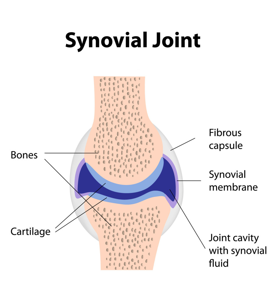
The knee is a synovial joint. Synovial joints have the most freedom to move. They're made of a cavity in one bone that another bone fits into. Slippery hyaline cartilage covers the ends of bones that make up a synovial joint. A synovial membrane — a fluid-filled sac that lubricates and protects the joint — lines the space between the bones.
Labelled Diagram Of Synovial Joint
A synovial joint, also known as diarthrosis, joins bones or cartilage with a fibrous joint capsule that is continuous with the periosteum of the joined bones, constitutes the outer boundary of a synovial cavity, and surrounds the bones' articulating surfaces. This joint unites long bones and permits free bone movement and greater mobility. [1]
Structure and function of synovial joints HSC PDHPE
GCSE AQA Skeletal system - AQA Synovial joints The skeleton is the central structure of the body and is made up of bones, joints and cartilage. The skeleton provides the framework for muscles.
An In Depth Look at the Bones Classification and Structure of Skeletal Joints Interactive
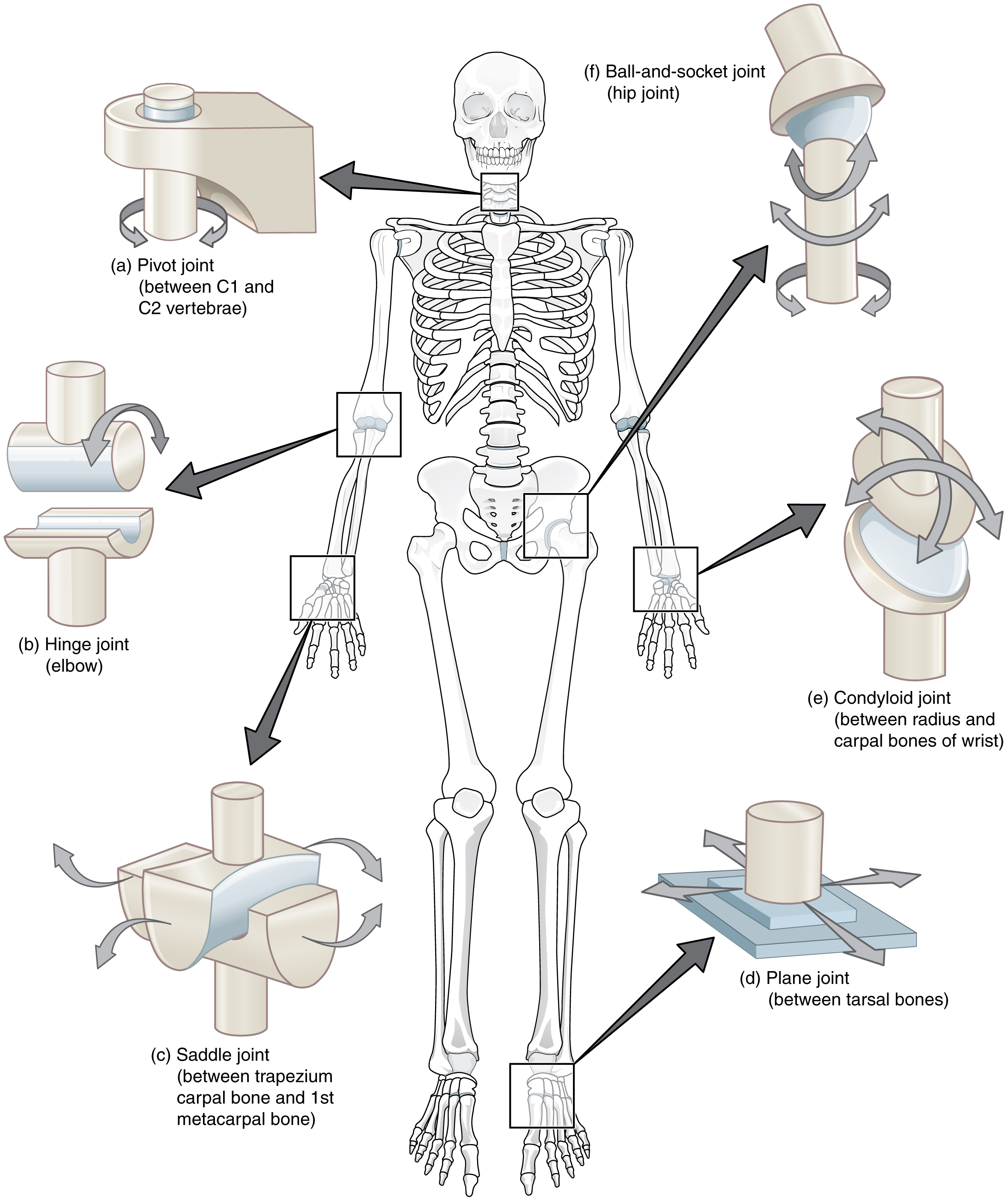
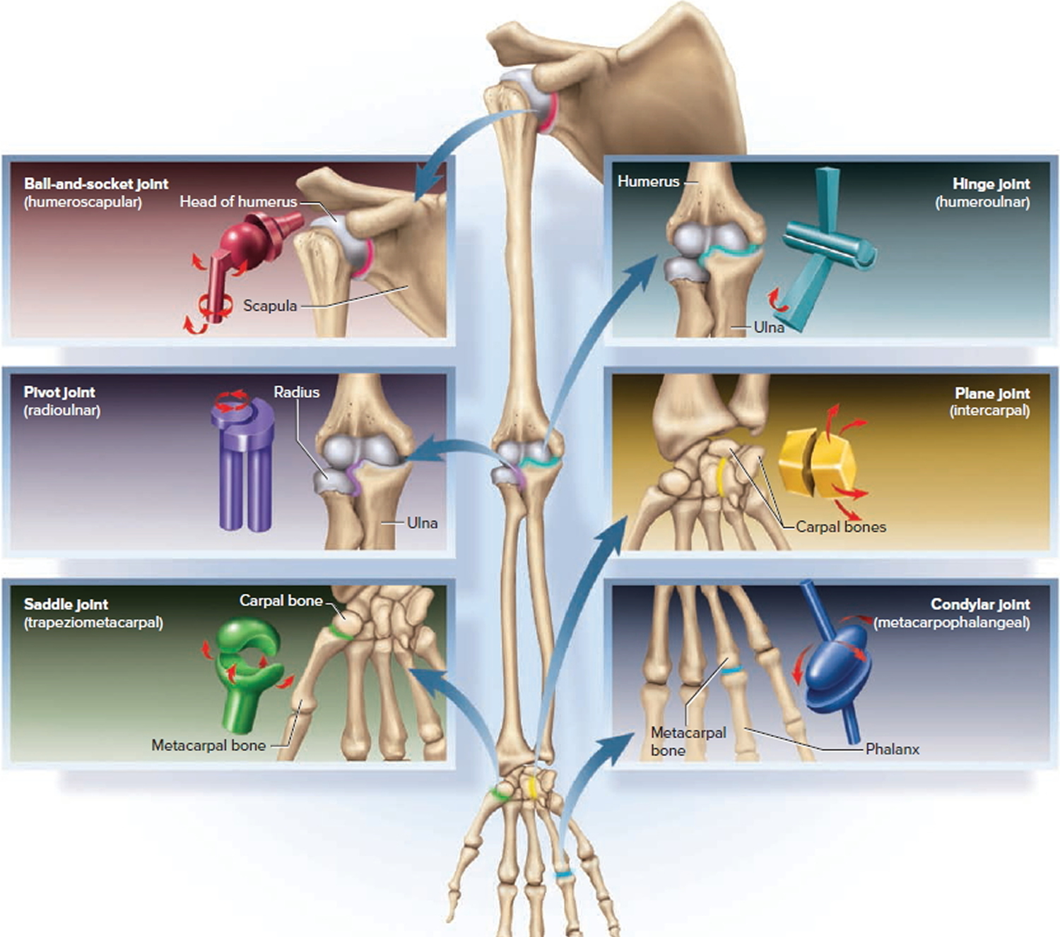
Figure 1. Different types of joints allow different types of movement. Planar, hinge, pivot, condyloid, saddle, and ball-and-socket are all types of synovial joints. Planar Joints Planar joints have bones with articulating surfaces that are flat or slightly curved faces.

Describe the structure of synovial joint with the
Figure 38.12.1 38.12. 1: Types of synovial joints: The six types of synovial joints allow the body to move in a variety of ways. (a) Pivot joints allow for rotation around an axis, such as between the first and second cervical vertebrae, which allows for side-to-side rotation of the head. (b) The hinge joint of the elbow works like a door hinge.

Structure of synovial joint English for Physio
A synovial joint is a connection between two bones consisting of a cartilage lined cavity filled with fluid, which is known as a diarthrosis joint. Diarthrosis joints are the most flexible type of joint between bones, because the bones are not physically connected and can move more freely in relation to each other.
Synovial Joint Structure
This is a pivot joint that allows for rotation of the radius during supination and pronation of the forearm. Figure 9.6.4 - Elbow Joint: (a) The elbow is a hinge joint that allows only for flexion and extension of the forearm. (b) It is supported by the ulnar and radial collateral ligaments.

9.4 Synovial Joints Anatomy & Physiology
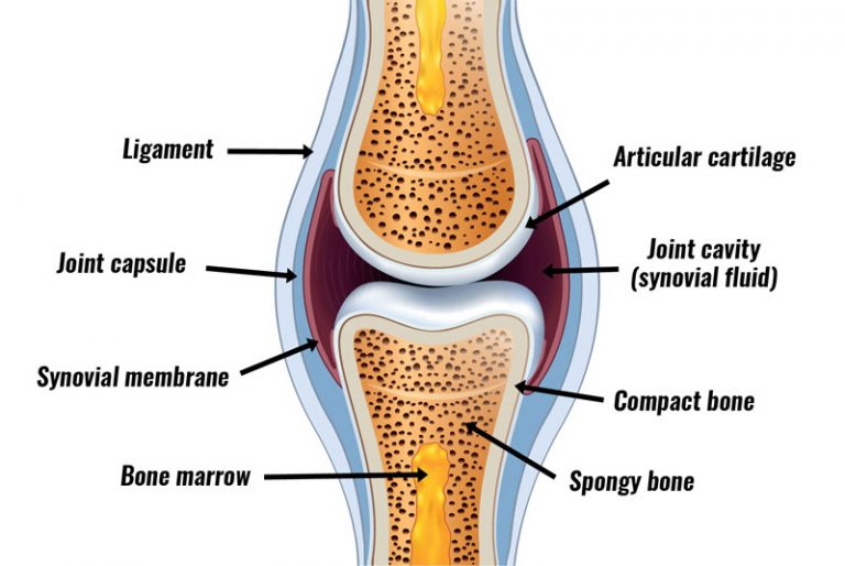
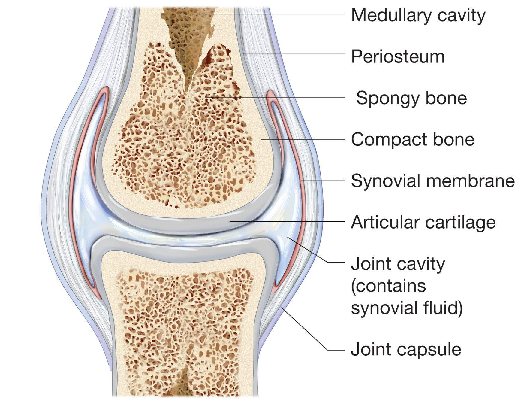
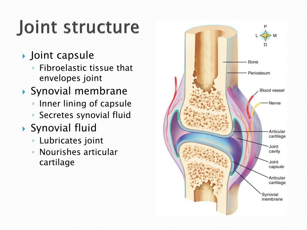
The cells of this membrane secrete synovial fluid (synovia = "a thick fluid"), a thick, slimy fluid that provides lubrication to further reduce friction between the bones of the joint. This fluid also provides nourishment to the articular cartilage, which does not contain blood vessels.

Synovial Joints Physiopedia
Synovial joints are characterized by the presence of a joint cavity. The walls of this space are formed by the articular capsule, a fibrous connective tissue structure that is attached to each bone just outside the area of the bone's articulating surface. The bones of the joint articulate with each other within the joint cavity.
Structure and function of synovial joints HSC PDHPE
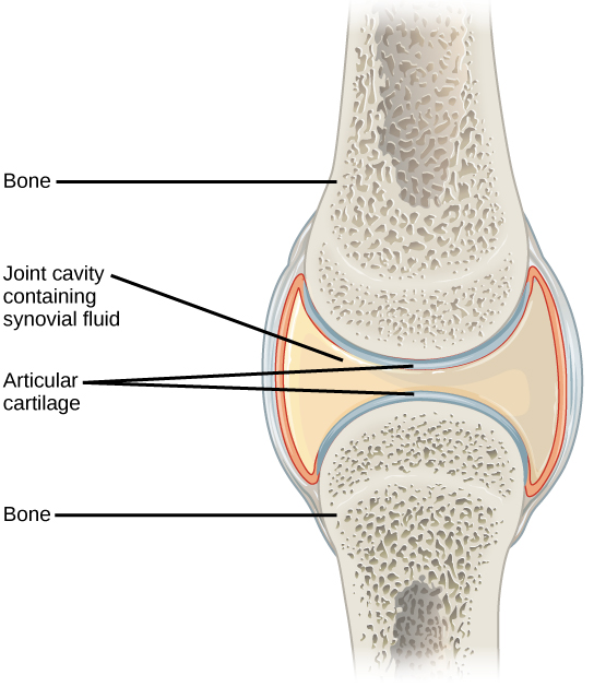
Synovial joints are the most common type of joint in the body (Figure 8.5.1 8.5. 1 ). A key structural characteristic for a synovial joint that is not seen at fibrous or cartilaginous joints is the presence of a joint cavity. This fluid-filled space is the site at which the articulating surfaces of the bones contact each other.

Human synovial joint Download Scientific Diagram
The articulating surfaces of a synovial joint (i.e. the surfaces that directly contact each other as the bones move) are covered by a thin layer of hyaline cartilage. The articular cartilage has two main roles: (i) minimising friction upon joint movement, and (ii) absorbing shock. Synovial Fluid

A general synovial joint. Download Scientific Diagram
What are synovial joints? Synovial joints have the most freedom to move. They're made of a cavity in one bone that another bone fits into. Slippery hyaline cartilage covers the ends of bones that make up a synovial joint. A synovial membrane — a fluid-filled sac that lubricates and protects the joint — lines the space between the bones.
Joints and Skeletal Movement · Biology
Synovial joints are the most common type of joint in the body (see image 1). These joints are termed diarthroses, meaning they are freely mobile. [1] A key structural characteristic for a synovial joint that is not seen at fibrous or cartilaginous joints is the presence of a joint cavity.

The Synovium, Synovitis, Inflammation, and Joint Pain HealDove
Synovial joints are the freely mobile joints in which the articulating surfaces have no direct contact with each other.The movement range is defined (i.e., limited) by the joint capsule, supporting ligaments and muscles that cross the joint. Most of the upper and lower limb joints are synovial.. The majority of the synovial joints are lined with hyaline cartilage, except for the.
PPT Structure of Synovial Joints PowerPoint Presentation, free download ID2504513
Synovial joints are characterized by the presence of a joint cavity. The walls of this space are formed by the articular capsule, a fibrous connective tissue structure that is attached to each bone just outside the area of the bone's articulating surface. The bones of the joint articulate with each other within the joint cavity.
Types & Classification of Body Joints Cartilaginous & Synovial Joint
A synovial joint, also known as a diarthrosis, is the most common and most movable type of joint in a mammal's body. Diarthroses are freely movable articulations. In these joints, the contiguous bony surfaces are covered with articular cartilage and connected by ligaments lined by synovial membrane. The joint may be divided, completely or.
